Your face, the one thing you scrutinize every morning under bathroom lighting — may Actually-explains”>Actually be doing more diagnostic work than you realize. Long before a physician orders a liver panel, long before numbers on a lab report tell any story, your skin can start to whisper something is wrong. Dermatologists and hepatologists have known this for decades. The rest of us, not so much.
The skin is a major target organ for extrahepatic manifestations of liver diseases, and clinical examination of the skin, nails, and hair can allow for appropriate recognition, early diagnosis, and treatment of liver disease, improving both quality of life and life expectancy. Think of your face as a dashboard, not just a canvas. Three specific areas stand out as the most telling, and, frankly, the most overlooked.
Key takeaways
- Tiny red spiders blooming across your cheeks and chest may point to something far more serious than aging
- Those subtle yellow patches near your eyes could be your skin’s urgent memo about cholesterol and liver function
- The whites of your eyes turn yellow before your skin does—and that timing window is critical for diagnosis
1. The Cheeks and Upper Chest: Spider Angiomas
Imagine a tiny red dot with delicate red lines radiating outward from it, like a miniature spider sitting just below the surface of your skin. That is a spider angioma, and one of them, on its own, is usually harmless. The plot thickens when they multiply.
Having more than three spider angiomas is likely to be abnormal and may be a sign of liver disease and/or hepatitis C; it also suggests the probability of esophageal varices. The mechanics behind this are worth understanding: individuals with significant liver disease also show many spider angiomas because their liver cannot metabolize circulating estrogens, specifically estrone, which derives from the androgen androstenedione. The liver loses its grip on hormone regulation, estrogen accumulates, blood vessels dilate, and the result blooms on your skin.
Spider angiomas are characteristically found on the face, neck, upper chest, and arms in adults, corresponding to the distribution of the superior vena cava. Multiple spider nevi usually indicate the presence of progressive hepatic fibrosis and advanced liver disease. Here’s the counter-intuitive part: these lesions are sometimes mistakenly attributed to rosacea, sun damage, or aging. A dermatologist can tell the difference in seconds, the classic spider angioma blanches completely when you press its center and then refills from the inside out when you release. No other vascular lesion behaves quite that way.
Non-specific signs such as spider angiomas, palmar erythema, jaundice, and pruritus may precede more overt clinical symptoms and offer valuable diagnostic and prognostic insights. So if you’ve noticed a cluster of these appearing where you never had them before, and you’re not pregnant or on hormonal contraception, that’s worth a conversation with your doctor, before any blood test has had the chance to catch up.
2. The Eyelids: Xanthelasma
Soft, flat, yellowish patches at the inner corners of the eyelids. They look almost polite, barely raised, subtle in color, easy to dismiss as a weird puffiness or the beginning of an allergy. They are, in fact, cholesterol deposits sitting just beneath your skin, and they carry a medical message your liver may already be sending.
Xanthelasma manifests as pale yellow, planar or slightly bulged, soft plaques around the eyelids, being essentially subcutaneous lipid deposits. The condition is associated with dyslipoproteinemia secondary to liver diseases such as primary biliary cholangitis (PBC) and other forms of cholestatic liver disease.
The connection to liver function runs deeper than simple cholesterol excess. Approximately half of patients with xanthelasma have underlying conditions causing hyperlipidemia and should be evaluated for underlying causes. Primary biliary cholangitis (PBC) is one known cause of xanthelasma and is an autoimmune condition that contributes to the destruction of bile ducts, leading to cholestasis. When the liver’s bile ducts are compromised, lipids back up into circulation, and the skin, particularly the delicate tissue around the eyes, becomes a storage site.
A xanthelasma diagnosis doesn’t require tests, but your healthcare provider may want to check your cholesterol levels, thyroid function, blood sugar, and liver function to find out if you have liver disease. The eyebrow-raiser here, pun intended, is that a xanthelasma is sometimes a symptom of a more serious underlying condition, such as dyslipidemia, hypothyroidism, kidney disease, liver disease, or diabetes. The eyelid patch that you’ve been meaning to ask about for the past year might be the skin’s version of a polite but urgent memo.
The condition is seen most frequently in females over 50 years old, and half of the cases present with comorbid dyslipidemia. Middle-aged and older women, in particular, should not brush these off as cosmetic nuisances.
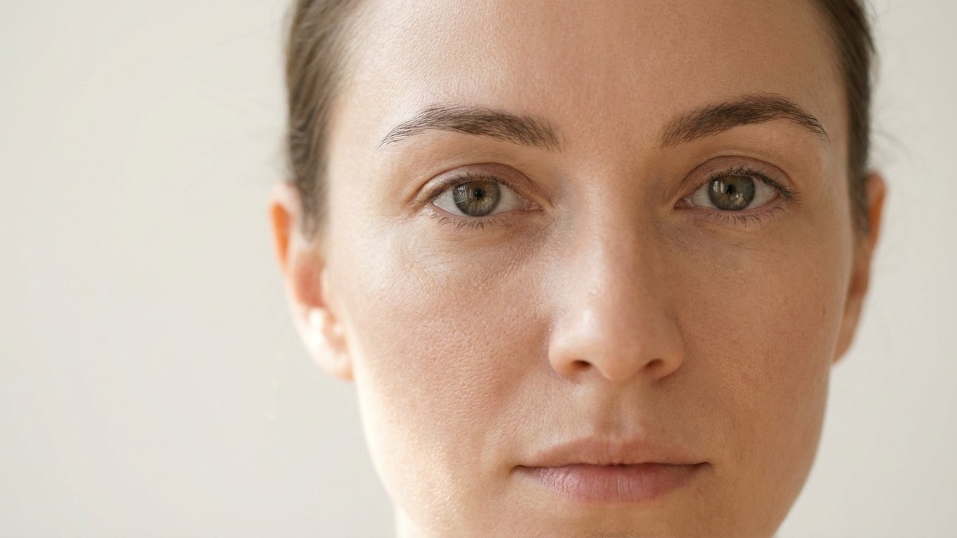
3. The Whites of the Eyes (and the Surrounding Skin): Scleral Icterus
This one is the face’s most direct alarm system. Scleral icterus is when the whites of your eyes look yellow. It’s a key early symptom of disruptions in your liver function. The reason: a failing liver cannot properly process bilirubin, the yellow pigment produced by the breakdown of red blood cells. That bilirubin has to go somewhere, and the eyes, specifically their conjunctiva, absorb it first.
The eyes are often the first location to take on a yellowish color when bilirubin levels get too high. Yellowing of the eyes is noticeable at bilirubin levels of 2–3 mg/dL, while yellowing of skin becomes noticeable at 4–5 mg/dL. This is the critical detail that most people don’t know: by the time the skin looks yellow, liver dysfunction has already progressed further. The eyes give you earlier warning. The window between eye yellowing and skin yellowing is precious diagnostic time.
Scleral icterus is most often the initial presenting sign of jaundice, which can be the result of alcohol-induced liver cirrhosis. This yellowing is often one of the first visible signs of a deeper problem, such as liver disease or hemolytic anemia. While it can appear subtle at first, any degree of scleral yellowing should be taken seriously.
The subtlety is the challenge. A very faint yellow cast in natural light, easily rationalized away as fatigue or poor lighting. Dermatologists recommend checking in daylight, against a white background, the contrast makes early tinting far more visible.
When to Act on What Your Face Is Telling You
Cirrhosis silently damages the liver, but its warning signs often appear on the skin first. Understanding these symptoms of liver cirrhosis can help detect liver disease early, leading to better outcomes. The three signs above, spider angiomas clustering on the face and chest, xanthelasma forming at the eyelid corners, yellowing of the eye whites — are not a diagnosis. They’re a prompt. A reason to call your doctor this week, not next month.
Given their accessibility during physical examination, these dermatologic features should be routinely assessed in patients at risk for chronic liver disease. Regular skin evaluations can facilitate earlier detection of liver cirrhosis, allowing for timely diagnostic workup, initiation of management strategies, and improved patient outcomes.
The irony, of course, is that we spend enormous energy analyzing what our skin looks like for cosmetic reasons, texture, pores, hyperpigmentation, but rarely consider what it might actually be communicating about the organs beneath it. Your face is not just an aesthetic surface. It’s a medical document. The question is whether you’re reading it.